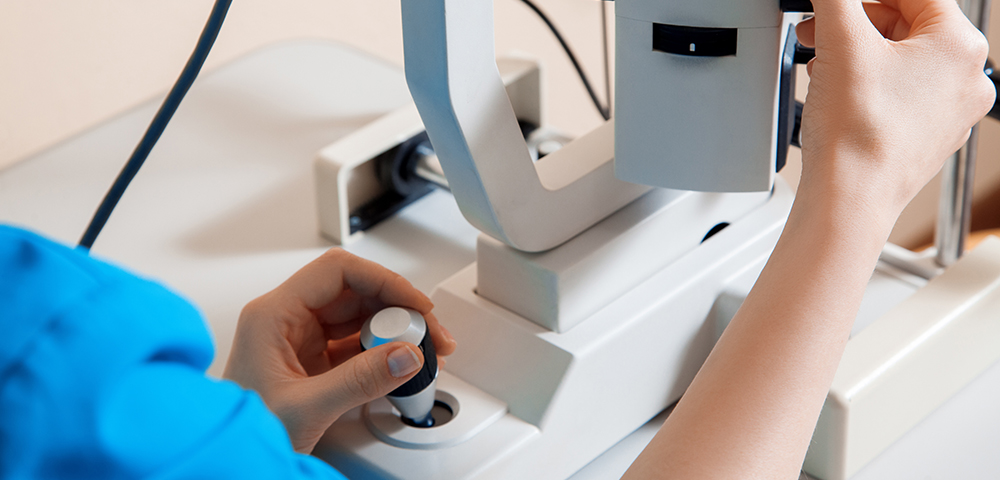
Fundus photography
Fundus photography involves a retinal camera designed to photograph the back and interior surface of the eye.
An optometrist may order fundus photography to diagnose or treat the following eye diseases:
- Glaucoma
- Diabetic retinopathy, such as Macular Edema and Microaneurysms
- Hypertensive retinopathy, which is an eye complication in people with high blood pressure
- Optic atrophy, which is eye nerve damage
- Papilledema, which is the swelling of the eye nerve
- Eye cancer
- Retinoblastoma, which is a tumour inside the eye
- Colour vision deficiencies
- Congenital glaucoma
- Congenital rubella
- Congenital anomalies
There are several benefits to fundus photography, some of which include:
- It is easier to see the retina’s details with fundus photography than directly looking at the eye.
- It provides a bird’s eye view of the layers on the retina so that an eye doctor can make an accurate diagnosis.
- It allows for early and accurate detection of eye diseases, especially in diabetes or high blood pressure patie
